NEUROLOGICAL TREATMENTS
COMMON TREATMENTS
The information outlined below on neurological treatments and specialist techniques is provided as a guide only and it is not intended to be comprehensive. Discussion with Mr Davies is important to answer any questions that you may have. For information about any additional diagnostics or treatments not featured within the site, please contact us for more information.
In anterior cervical discectomy with fusion, the surgeon approaches the cervical spine through a small incision in the front of the neck and removes the total disc or a part of the disc along with any bony material that is compressing or putting pressure on the nerves and producing pain. Spinal fusion implies placing a bone graft between the two affected vertebral bodies encouraging the bone growth between the vertebrae. The bone graft acts as a medium for binding the two vertebral bones, and grows as a single vertebra that stabilises the spine. It also helps to maintain the normal disc height.
As most nerves to the body (e.g., arms, chest, abdomen, and legs) pass through the neck region from the brain, pressure on the spinal cord in the neck region (cervical spine) can be very problematic. Patients with these symptoms are potential candidates for anterior cervical discectomy procedure but only after non-surgical treatment methods fail. Cervical discectomy can reduce the pressure on the nerve roots and provide pain relief.
The Procedure
Your surgeon makes a small incision in the front side of the neck and locates the source of neural compression (pressure zone). Then, the intervertebral disc that is compressing the nerve root will be removed. Afterwards, a bone graft will be placed between the two vertebral bodies. In certain instances, metal plates or pins may be used for providing enough support and stability, and to ease the fusion of the vertebrae.
Postoperative Instructions
A specific post-operative recovery/exercise plan will be given by your physician to help you return to normal activity at the earliest possible. The duration of hospital stay depends on this treatment plan. You will be able to wake up and walk by the end of the first day after the surgery. You would be able to resume your work within 3-6 weeks, depending on your body’s healing status and the type of work/activity that you plan to resume. Discuss with your spinal surgeon and follow the instructions for optimised healing and appropriate recovery after the procedure.
Risks and Complications
Treatment results are different for each patient. In addition to the anaesthetic complications, spinal surgery is associated with some potential risks such as infection, blood loss, blood clots, nerve damage, and bowel and bladder problems. Failure to fuse the vertebral bones with the bone graft (fusion failure) is an important complication of spinal fusion which requires an additional surgery.
Please take your physician’s advice for a complete list of indications, clinical results, adverse effects, warnings and precautions, and other relevant medical information about anterior cervical discectomy with fusion surgery.
Some biopsies are minimally invasive. They involve a needle and imaging guidance to target and collect tissue samples. Others require open access to the brain tissue through the skull.
Types of Brain Biopsy
There are two primary types of brain biopsies:
- Stereotactic needle biopsy: This minimally invasive approach uses precise imaging guidance to target and collect a tissue sample from the brain. Doctors may prefer it for lesions that occur deep within the brain. Doctors most oftenTrusted Source use this type of biopsy technique.
- Open brain biopsy: In this procedure, a neurosurgeon creates a small opening in the skull to access the brain tissue directly. They may use it for more extensive tissue sampling or when the lesion’s location makes stereotactic biopsy challenging.
The Procedure
The following occurs during a brain biopsy:
- Anesthesia administration: This is where a person receives either local or general anesthesia. This helps ensure comfort and minimizes any discomfort or pain during the procedure.
- Stereotactic needle biopsy technique: Doctors use precise and advanced imaging techniques to guide a thin, hollow needle to the target area within the brain tissue.
- Open brain biopsy: Alternatively, for an open brain biopsy, a neurosurgeon temporarily removes a small section of the skull. This opening allows direct access to the brain tissue that requires examination.
- Tissue collection and analysis: The doctor carefully collects a brain tissue sample. The team then promptly sends the tissue sample to a laboratory for thorough analysis to determine if there are any neurological conditions or issues.
After collecting the brain tissue sample, a pathologist conducts a thorough examination. The results provide detailed insights into the characteristics of the cells, including if they are benign or malignant, infectious, or inflammatory. This information helps doctors shape treatment strategies and clinical decisions.
A brain biopsy is a diagnostic procedure doctors use to obtain brain tissue for examination. Most people have them as they are less invasive and involve a faster recovery. The other type of biopsy involves accessing the brain through a hole that doctors cut in the skull.
Both types of biopsy are safe procedures that provide doctors with critical information they cannot determine from imaging studies alone.
Disorders that can affect the craniocervical junction later in life include:
- Rheumatoid arthritis
- Paget’s disease
- Tumors of the craniocervical junction
Disorders that can affect the craniocervical junction from birth can involve isolated or general disorders.
Isolated disorders include:
- Platybascraniocervical junction surgeryia
- Basilar invagination
- Atlas assimilation
- Atlantoaxial subluxation or dislocation
- Atlas hypoplasia
- Klippel-Feil malformation
- Chiari malformation
- Os odontoideum
General disorders may include the same abnormalities in the craniocervical junction as isolated disorders, but these are part of a larger group of abnormalities, such as:
- Achondroplasia
- Down syndrome
- Mucopolysaccharidosis
- Osteogenesis imperfecta
Craniocervical Junction Surgery
Craniocervical junction surgery is an operation performed on the bones in the junction between the skull and the spine. The craniocervical junction includes the bone that forms the base of the skull, called the occipital bone, and the first two bones in the spine, called the atlas and the axis.
Craniocervical junction surgery may be used to treat disorders or deformities of the upper neck that are present at birth or occur later in life.
Disorders that affect the large opening at the bottom of the occipital bone (called the foramen magnum) are a particular concern because important structures pass through this opening. These structures include the lowest part of the brain (brain stem), which transitions into and is structurally continuous with the spinal cord, as well as certain nerves and blood vessels.
If your headaches are severe or you have problems caused by the pressure on your spinal cord (such as movement difficulties), surgery may be recommended.
Chiari Compression Surgery
The main operation for Chiari malformation is called decompression surgery.
Under general anaesthetic a cut is made at the back of your head and the surgeon removes a small piece of bone from the base of your skull. They may also remove a small piece of bone from the top of your spine.
This will help reduce the pressure on your brain and allow the fluid in and around your brain and spinal cord to flow normally. Other procedures that may be necessary include:
- Endoscopic third ventriculostomy (ETV) – a small hole is made in the wall of one of the cavities of the brain, releasing trapped fluid. See treating hydrocephalus for more information.
- Ventriculoperitoneal shunting – a small hole is drilled into the skull and a thin tube called a catheter is passed into the brain cavity to drain trapped fluid and relieve the pressure. See treating hydrocephalus for more information.
- Untethering – some children with a type 1 Chiari malformation have a tethered spinal cord, which means it is abnormally attached within the spine. Untethering involves separating the spinal cord and releasing tension in the spine.
- Spinal fixation – some people with Chiari I will have a hypermobility syndrome, such as Ehlers-Danlos syndrome, and may require surgery to stabilise their spine.
The aim of surgery is to stop existing symptoms getting any worse. Some people also experience an improvement in their symptoms, particularly their headaches.
However, surgery sometimes results in no improvement or symptoms getting worse. There’s also a small risk of serious complications, such as paralysis or a stroke. Talk to your surgeon about the different surgical options and what the benefits and risks of each are.
Cranioplasty is the surgical repair of a bone defect in the skull resulting from a previous operation or injury. There are different kinds of cranioplasties, but most involve lifting the scalp and restoring the contour of the skull with the original skull piece or a custom contoured graft made from material such as:
- Titanium (plate or mesh).
- Synthetic bone substitute (in liquid form).
- Solid biomaterial (prefabricated customized implant to match the exact contour and shape of the skull).
Conventional cranioplasty methods, which have been used by neurosurgeons for more than 100 years, involve peeling back all five layers of the scalp to place the bone remnant or custom implant into the proper cranial location. For the pericranial-onlay cranioplasty, a newer technique developed here at Johns Hopkins by Chad Gordon and his team, the surgeon gently pulls back only the three uppermost layers of the scalp and inserts the bone or implant in between the bottom layers of the scalp protecting the brain. This type of cranioplasty procedure is safer and less invasive.
Why you might need a Cranioplasty
Cranioplasty might be performed for any of the following reasons:
- Protection: In certain places, a cranial defect can leave the brain vulnerable to damage.
- Function: Cranioplasty may improve neurological function for some patients. In some instances, a customized cranial implant is designed ahead of time to help the surgeon obtain an ideal shape and outcome, as well as to house embedded neuro technologies.
- Aesthetics: A noticeable skull defect can affect a patient’s appearance and confidence.
- Headaches: Cranioplasty can reduce headaches due to previous surgery or injury.
The Procedure
In the operating room, you are given a general anesthetic. Once you are asleep, the team positions you so the surgeons have optimal access to the bone defect. The area of the incision is then shaved and prepared with antiseptic, and you are protected by drapes that leave only the surgical area exposed.
You will get a local anesthetic, then the surgeon will carefully cut the skin of your scalp and gently separate it into layers, thereby protecting the dura, which covers the brain. The team cleans the edges of surrounding bone and prepares the surface so the bone or implant can be positioned properly in the defect, after which it is secured to the cranial bones with screws, plates or both.
With the bone or implant in place, bleeding is controlled, the team moves the scalp back to its original position and closes the incision with nylon suture. You may also have a small suction drain left in place to help remove any excess fluid. The drain will be removed in a few days.
Post Operative Recovery
You will wake up in recovery, and after about an hour you will be transferred to the neurosurgical floor or to the NCCU (neurosurgical intensive care unit). Your nursing staff will continually monitor you for any signs of a complication, and measure your pulse, blood pressure, limb strength and level of alertness. During the first night in the hospital, you will be awakened for these observations.
Operations on the head do not often hurt much, but you may have a headache and will have pain relief pills and injections to ensure you’re comfortable. You may still have a urinary catheter in place from the operation.
In the next day or so, your nurse will remove the IV drip in your arm, and you will be encouraged to walk. Gradually, you will be able to move about normally. Your head bandage will be removed on the second day after surgery.
Most cranioplasty patients spend two to three days in the hospital after surgery. When your care team determines you can get around, shower and dress yourself, you will get a repeat CT scan of your head. If the surgical site looks okay, you will be released and can go home.
A craniotomy is an operation to open the head in order to expose the brain. The word craniotomy means making a hole (-otomy) in the skull (cranium).
What is a Craniotomy used for?
A craniotomy is necessary to deal surgically with a number of abnormalities of the brain and its surrounding structures. The following are a few examples of the types of condition for which a craniotomy is commonly carried out.
- Severe head injury, which results in a blood clot pressing on the brain. If the blood clot is formed between the membranes surrounding the brain, it is known as a subdural haematoma. If the blood clot is between the inside of the skull and the outer membrane covering the brain, it is known as an extradural haematoma.
- Growth or tumour: a tumour can develop either from the membranes surrounding the brain (for example, a meningioma) or within the brain itself (for example, a glioma). Any such growth can cause pressure on the brain.
- Bleeding in or on the brain: This can happen due to abnormal blood vessel leakage, leading to conditions like a subarachnoid haemorrhage.
The Procedure
The operation is carried out under a general anaesthetic. You will be asleep and will not feel anything. It will be necessary to shave a small section of the head. The place, size and shape of the skin cut (incision) vary according to the type of operation.
The incision is usually placed behind the hairline to hide the scar although this is not always possible. The scar will fade to a pale thin line within 3 to 6 months and the hair will usually grow back normally where it has been shaved.
To reach the brain, a small part of the skull is temporarily removed. The exact location of the opening is decided after careful consideration of brain scans and other investigations that have been carried out before the operation.
Once the opening has been made, the lesion (abnormal tissue or growth) is then removed or treated.
After the surgery has been completed, the bone is then replaced to cover the hole that has been made.
Recovery from Craniotomy
Recovery time depends on the underlying condition and on whether there were any complications during or after the operation. Normally, you can expect to stay in hospital for 5 to 10 days and rest at home for a further 6 to 12 weeks.For people who have problems related to their underlying condition, there may be a need for a longer hospital stay and further care in a rehabilitation unit.
Types of Epilepsy Surgery
- Resective surgery. This is the most common epilepsy surgery. The surgeon removes a small portion of brain tissue from the area of the brain where seizures occur. Resective surgery is usually performed on one of the temporal lobes, an area of the brain that controls emotions, visual memory, and understanding language.
- Disconnection surgery. This is where the surgeon disconnects one part of the brain from another part to stop the seizure from spreading. This is done either by cutting the connections between the two halves (hemispheres) of the brain. Or it can be done by cutting the nerve fibres in the outer layers of the brain.
- Laser Interstitial Thermal Therapy (LITT). This is also known as laser beam therapy. It is different from the other surgeries as the wound left afterwards is very small, which means that, often, you can go home the next day. With LITT a laser is used to pinpoint and destroy a small part of the brain that is causing the seizures. It is carried out in a Magnetic Resonance Imaging (MRI) scanner.
Pre-Operative Tests
If you are referred for surgery assessment you will probably go to a specialist centre for tests. There are many different pre-surgical tests you might have before you can be given the go-ahead for surgery. These can include MRI scans, an EEG (electroencephalogram) and video telemetry (an EEG while also being filmed). Other types of scan may also be done, which trace a chemical injected into the body. This can show detailed information about where seizures start in the brain.
Memory and psychological tests are also used to see how your memory and lifestyle might be affected after the surgery. These types of tests also help the doctors to see how you are likely to cope with the impact of having this type of surgery. The tests will confirm whether:
- the surgeons can reach the epileptogenic lesion during surgery and can remove it safely without causing new problems;
- other parts of your brain could be affected by the surgery, for example the parts that control your speech, sight, movement, memory, or hearing;
- you have a good chance of having your seizures stopped by the surgery and better quality of life; and
- you have any other medical conditions that would stop you from having this kind of surgery.
The results from the pre-surgical tests will help you and your neurologist decide whether surgery is an option for you, and what the result of the surgery might be. Your specialist will also talk with you about the possible risks and benefits of having surgery.
Surgery and Recovery
The most common type of epilepsy surgery is removal of part of the temporal lobe. Possible risks of this type of surgery include problems with memory, a partial loss of sight, depression, or other mood problems. These risks will vary from person to person, and may be only temporary in some cases. For some people, their memory and mood could improve after epilepsy surgery.
Immediately after the surgery your doctors will monitor your recovery. For the first few days you may feel very tired and need to sleep, as it can take a while for the anaesthetic to completely wear off. Some people who have brain surgery will have seizures within the first week of surgery – but this does not mean the surgery has not been successful.
Seizures after surgery can happen because of the direct stress the brain experiences in surgery. How long you may need to spend in hospital will depend on the type of surgery you have had and how you are recovering. Generally your doctors might expect you to be back to your normal activities about six weeks after your surgery, but this is very individual.
Around 70% of people (7 in 10 people) who have temporal lobe surgery find that the surgery stops their seizures and they become seizure-free, and for a further 20% (1 in 5 people) their seizures are reduced. Around 50% of people (half) who have temporal lobe surgery are still seizure-free 10 years after their surgery, but most of these people will still take their ASM for some time.
During shunt surgery, a thin tube called a shunt is implanted in your brain. The excess cerebrospinal fluid (CSF) in the brain flows through the shunt to another part of your body, usually your tummy. From here, it’s absorbed into your blood.
Inside the shunt there’s a valve that controls the flow of CSF, so it does not drain too quickly. You can feel the valve as a lump under the skin on your scalp.
The Operation
Shunt surgery is done by a specialist in brain and nervous system surgery (neurosurgeon). It’s done under a general anaesthetic and usually takes 1 to 2 hours. You may need to stay in hospital for a few days after the operation to recover.
If you have stitches, they may dissolve or need to be removed. Some surgeons use skin staples to close the wound, which will need to be removed after a few days. After the shunt has been installed, further treatment for hydrocephalus may be needed if the shunt becomes blocked or infected. Shunt repair surgery will then be necessary.
Endoscopic third ventriculostomy (ETV)
An alternative procedure to shunt surgery is an endoscopic third ventriculostomy (ETV). Instead of inserting a shunt, the surgeon makes a hole in the floor of your brain to allow the trapped cerebrospinal fluid (CSF) to escape to the brain’s surface, where it can be absorbed.
ETV is not suitable for everyone, but it could be an option if the build-up of CSF in your brain is caused by a blockage (obstructive hydrocephalus). The CSF will be able to drain through the hole, avoiding the blockage.
THE OPERATION
ETV is done under general anaesthetic. The neurosurgeon makes a small hole in your skull and brain, and uses an endoscope to look inside the chambers of your brain. An endoscope is a long, thin tube with a light and camera at one end.
After making a small hole in the floor of your brain to drain the fluid, the endoscope is removed and the wound is closed using stitches. The procedure takes around 1 hour.
There’s less risk of infection after ETV than with shunt surgery. However, as with all surgical procedures, there are some risks.
The long-term results of ETV are similar to those for a shunt operation. As with shunts, there’s a risk of a blockage happening months or years after surgery, which will cause your symptoms to return.
SPECIALIST TECHNIQUES
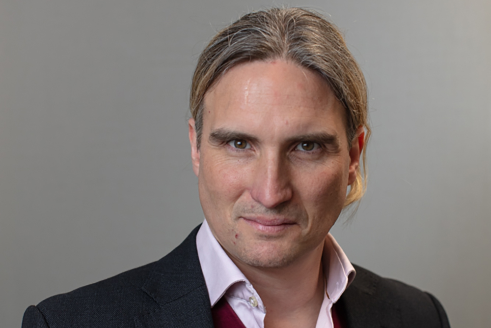
Mr David Davies holds specialist interests in the development of optical techniques within the context of traumatic brain injury, particularly working in conjunction with physical and computer scientists. He is very keen to incorporate contemporary research from the seemingly unrelated disciplines of engineering and computer sciences into the field of neurosurgical trauma. He is involved in many national and international trials into the treatment of a variety of operative and non-operative aspects of traumatic brain injury, and has experience in many specialist techniques including cerebral microdialysis, brain tissue oxygen monitoring and multi-modal MR imaging.
For more information please don’t hesitate to contact us at info@djdsurgery.co.uk and a member of our team will get back to you as soon as possible.


